Surgery as the standard of care to monitor head and neck cancer
The PET-NECK study set out to investigate if PET-CT scans could be used to monitor disease in patients after chemoradiotherapy, with neck surgery only used on those that still had disease or unclear results, rather than surgery as routine.
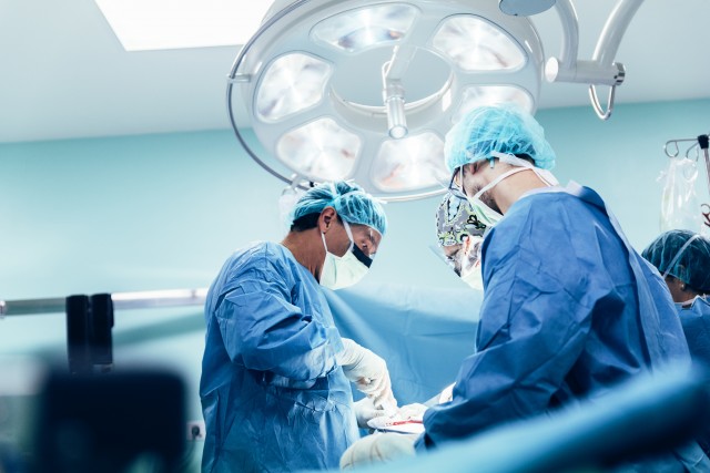
Traditionally it has been difficult to monitor the response of head and neck cancer to chemoradiotherapy using imaging scans. As a result, the standard care for patients with some kinds of head and neck cancer has included neck surgery after chemoradiotherapy. This is an invasive 2-3 hour operation and requires a hospital stay. It can be technically difficult for surgeons and carries risks of life-changing complications. However, other techniques to monitor the cancer in these patients were limited, and so surgery was necessary.
The NCRI Head & Neck Group identified this as an opportunity to transform the standard of care for this group of patients, in developing a less invasive way to monitor people with head and neck cancer. There was evidence from small retrospective studies that PET-CT scans could identify the absence of tumour in the neck following chemoradiotherapy, however there was a need for a definitive trial to provide the evidence needed to change clinical practice.
Proving coordination for PET-NECK through the NCRI Head & Neck Group
PET-NECK is the largest trial completed in head and neck cancer in the UK in the last two decades, with 564 patients recruited. The NCRI Head and Neck Group was at the centre of the development of the study.
Professor Hisham Mehanna, lead researcher on the study, explained how the Head & Neck Group supported the study: “The Group was essential in developing the idea for PET-NECK, applying for funding, running the study and ensuring and encouraging recruitment”.
“The study would have probably not happened if there had not been the input and coordination of the Group. Indeed, this Group is the envy of many head and neck cancer colleagues around the world”
The value of the NCRI Groups to studies like this is far-reaching; as explained by Professor Mehanna, the Group “originate new ideas, develop them, help others to develop their ideas, has significant expertise in obtaining grants, and is able to access the wider head and neck community and encourage recruitment and participation. This was my first major grant application and my first ever clinical trial – I needed help and could not have done it without the Group!”
Translating research into practice
The PET-NECK study successfully showed that PET-CT scans could be used as a monitoring technique, and that neck dissection surgery could be avoided in over 80% of patients*. This has great cost-saving potentials for the NHS – for each person that avoided neck dissection during the trial £1,492 was saved. The avoidance of invasive surgery and its potential complications is also a huge improvement in the care of patients, avoiding the risk of complications, reducing hospital stays and ensuring quicker recovery times.
This work has translated into new recommendations in clinical guidelines at a UK, European (e.g. Germany, Italy) and global level (US), with tangible changes seen in clinical practice in the UK and internationally. As a result of this study, PET-CT guided surveillance has now become standard care in the NHS. This research will benefit around 2,500 patients in the UK every year, estimates Professor Mehanna.
*The results of this study can be found in the publication: Mehanna, H et al. (2016). PET-CT surveillance vs neck dissection in advanced head and neck cancer. New England Journal of Medicine, 374;15 1444-1454.